Bone Health in Premenopausal Women: When to Screen and How to Treat
 Sam Anderson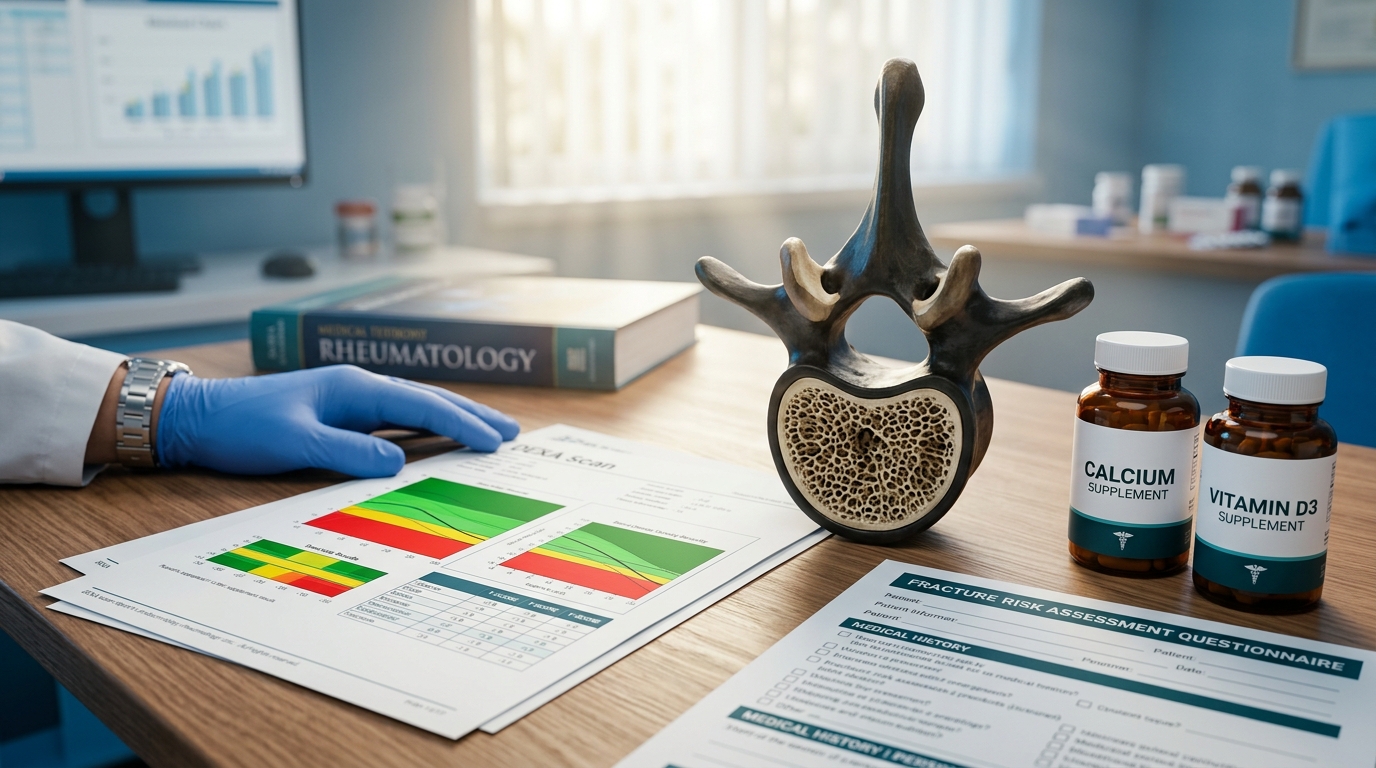
When to Screen Premenopausal Women
Bone health assessment in premenopausal women presents unique challenges that differ fundamentally from postmenopausal osteoporosis management. Standard DEXA interpretation using T-scores is not appropriate in premenopausal women — Z-scores are the correct metric — and the threshold for treatment is higher because many conditions causing low bone density in younger women are reversible with treatment of the underlying cause. For the endocrinologist, rheumatologist, or primary care physician evaluating a premenopausal woman with low bone density, the clinical approach must focus on identifying secondary causes before considering pharmacotherapy.
Unlike postmenopausal osteoporosis, universal screening is not recommended for premenopausal women. The ISCD (International Society for Clinical Densitometry) recommends DXA only in premenopausal women with fragility fractures, diseases associated with bone loss (e.g., celiac disease, hyperparathyroidism, anorexia nervosa, inflammatory bowel disease), or chronic medication use known to affect bone (glucocorticoids above 5 mg prednisone equivalent for 3 or more months, aromatase inhibitors used in breast cancer treatment, GnRH agonists, certain anticonvulsants). Critically, T-scores should not be used for premenopausal classification. Z-scores are appropriate, with a Z-score of -2.0 or below defined as low bone mineral density for age, not osteoporosis[1].
Secondary Cause Evaluation
When low BMD is identified in a premenopausal woman, a thorough secondary cause evaluation is essential. Standard workup includes comprehensive metabolic panel, phosphorus, 25-hydroxyvitamin D, PTH, TSH, celiac panel (tTG-IgA and total IgA), 24-hour urine calcium and creatinine, CBC, and ESR/CRP. Based on clinical suspicion, additional testing may include serum protein electrophoresis (multiple myeloma), cortisol testing (Cushing syndrome), prolactin and gonadotropins (hypothalamic amenorrhea), and bone turnover markers (CTX and P1NP). Secondary causes are identified in 50-70% of premenopausal women with unexplained low BMD[2].
Treatment: Addressing the Underlying Cause First
The primary treatment strategy is addressing reversible causes. For glucocorticoid-induced osteoporosis, the 2022 ACR guidelines recommend bisphosphonates for premenopausal women at moderate-to-high fracture risk receiving prednisone 2.5 mg or more daily for 3 or more months. Calcium intake of 1,000 mg daily and vitamin D supplementation to maintain 25(OH)D above 30 ng/mL are foundational. Weight-bearing exercise (150 minutes per week of moderate-intensity activity) increases BMD by 1-2% at the spine over 12 months in premenopausal women[2]. For hypothalamic amenorrhea, restoring menstrual function through energy balance correction or transdermal estradiol plus cyclic progesterone is the primary intervention.
Pharmacotherapy Considerations
Bisphosphonates are the most commonly used pharmacologic agents in premenopausal women at high fracture risk. Alendronate 70 mg weekly or risedronate 35 mg weekly increase lumbar spine BMD by 4-6% over 2 years in glucocorticoid-treated patients[2]. A critical consideration is the long skeletal half-life of bisphosphonates (up to 10 years for alendronate) and their teratogenic potential (FDA category X)[2]. Women of childbearing potential should be counseled about contraception and ideally have bisphosphonates discontinued 6-12 months before planned conception. Teriparatide (20 mcg daily) is an alternative for severe cases, with the advantage of shorter washout but higher cost and injection requirement.
Monitoring and Follow-Up
Repeat DXA is recommended at 1-2 year intervals, with the least significant change (LSC) for the specific DXA machine determining whether observed changes represent true biological change. Bone turnover markers (serum CTX for resorption, P1NP for formation) respond within 3-6 months of treatment initiation and can provide earlier evidence of treatment response. A decrease in CTX of 25% or more or P1NP of 30% or more from baseline suggests adequate antiresorptive effect. Transition planning to postmenopausal osteoporosis management should begin in perimenopause, recognizing that the accelerated bone loss phase (2-3% per year) occurs in the 2-3 years surrounding menopause[2].
The Key Clinical Distinction: Finding the Cause, Not Just the Number
The most important conceptual difference between premenopausal and postmenopausal bone management is this: in a postmenopausal woman, low bone density is expected and treatment is directed at the bone itself. In a premenopausal woman, low bone density is always a signal that something else is going on — and finding and treating that something else is the intervention most likely to improve bone outcomes. The endocrinologist who identifies celiac disease, corrects vitamin D deficiency, treats Cushing syndrome, or restores menstrual function in a patient with hypothalamic amenorrhea addresses the root cause rather than masking the consequence with bisphosphonate therapy. Pharmacotherapy has its place in premenopausal bone health, but it should follow — not replace — a thorough secondary cause evaluation.
Limitations and Practical Challenges
The evidence base for pharmacotherapy in premenopausal osteoporosis is thinner than for postmenopausal disease — fracture reduction trials in premenopausal women are limited, and treatment recommendations are extrapolated largely from glucocorticoid-induced osteoporosis data and postmenopausal studies. The teratogenicity concern with bisphosphonates creates real clinical tension when treating women who may wish to become pregnant in the future. And the psychological impact of a "low bone density" finding in a young woman should not be underestimated — careful communication about what Z-scores mean (and do not mean), the difference between low BMD and osteoporosis, and the excellent prognosis when underlying causes are identified and treated is essential for preventing unnecessary anxiety and overtreatment.
References
- ISCD 2005 Official Positions PubMed 16731428
- Evaluation of premenopausal osteoporosis PubMed 20485914
Frequently Asked Questions
Should T-scores or Z-scores be used for premenopausal women?
When is DXA screening indicated in premenopausal women?
How often are secondary causes identified in premenopausal low BMD?
Are bisphosphonates safe before planned pregnancy?
What bone turnover marker changes indicate treatment response?
How much does weight-bearing exercise increase BMD in premenopausal women?
Explore This Topic in Ailva
Ailva is a free clinical intelligence platform for NPI-verified US physicians. Get evidence-based answers with verified citations from 16M+ indexed papers — plus free CME credits.
Founder of Ailva.ai | Former Director of Research and Author of 200+ Medically Reviewed Articles | Editor-in-Chief of EudaLife Magazine