Thyroid Nodule Evaluation: Bethesda Classification, Molecular Testing, and When to Biopsy
 Sam Anderson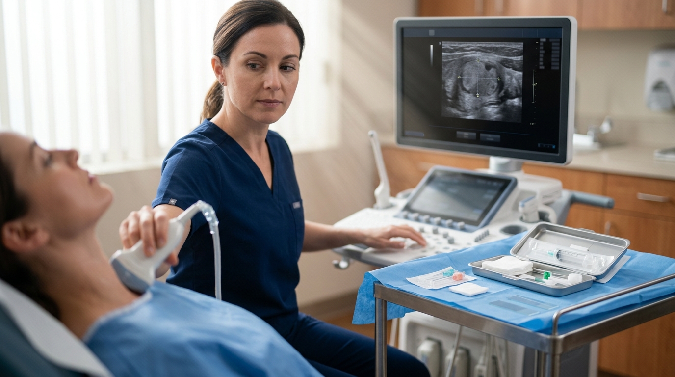
Ultrasound Risk Stratification: ACR TI-RADS
Thyroid nodules are extraordinarily common — found in up to two-thirds of adults when looked for with high-resolution ultrasound — yet the vast majority are benign. The clinical challenge is not finding thyroid nodules; it is determining which ones need further evaluation and which can be safely observed. Getting this triage wrong in either direction has consequences: missing a malignancy delays treatment, while over-investigating benign nodules subjects patients to unnecessary biopsies, surgeries, and anxiety. For the endocrinologist, radiologist, surgeon, or primary care physician managing thyroid nodules, the structured approach from ultrasound risk stratification through FNA to molecular testing provides a systematic framework that optimizes diagnostic accuracy while minimizing unnecessary procedures.
The ACR Thyroid Imaging Reporting and Data System (TI-RADS) assigns points based on five ultrasound categories: composition (0-2 points), echogenicity (0-3), shape (0-3), margin (0-3), and echogenic foci (0-3). Nodules scoring TR1 (benign, 0 points) or TR2 (not suspicious, 2 points) do not require FNA regardless of size. TR3 nodules (mildly suspicious, 3 points) warrant FNA at 2.5 cm or greater, TR4 (moderately suspicious, 4-6 points) at 1.5 cm, and TR5 (highly suspicious, 7+ points) at 1.0 cm. This system reduces unnecessary biopsies by approximately 30% compared to older ATA guidelines. For related screening approaches, see cancer screening guidelines[1].
Bethesda System for Reporting Thyroid Cytopathology
The Bethesda System (third edition, 2023) classifies FNA results into six categories with associated malignancy risks: Bethesda I (nondiagnostic, 5-10% malignancy risk), Bethesda II (benign, 0-3%), Bethesda III (atypia of undetermined significance/AUS, 10-30%), Bethesda IV (follicular neoplasm, 25-40%), Bethesda V (suspicious for malignancy, 50-75%), and Bethesda VI (malignant, 97-99%)[5]. The updated third edition revised the implied malignancy risk for Bethesda III upward, reflecting real-world data showing higher cancer rates than initially estimated.
For Bethesda I results, repeat FNA should be performed after 4-6 weeks. Bethesda II nodules undergo surveillance ultrasound at 12-24 months. Bethesda V and VI typically proceed to surgery. The clinical dilemma concentrates on Bethesda III and IV, where molecular testing has become the critical decision tool.
Molecular Testing in Indeterminate Nodules
The two dominant commercial platforms are the Afirma Genomic Sequencing Classifier (GSC) and ThyroSeq v3. Afirma GSC functions primarily as a rule-out test with a negative predictive value of 96% for Bethesda III and 95% for Bethesda IV nodules[2]. A benign Afirma result supports observation over surgery. ThyroSeq v3 analyzes 112 genes and functions as both a rule-in and rule-out test, with sensitivity of 94% and specificity of 82% in the indeterminate cytology population[3]. A negative ThyroSeq result carries a residual cancer risk of approximately 3%.
Choosing Between Afirma and ThyroSeq in Practice
The choice between Afirma GSC and ThyroSeq v3 for indeterminate nodules often depends on institutional preference and what clinical question you are trying to answer. If your primary goal is to rule out malignancy and avoid surgery — the most common clinical scenario for Bethesda III nodules — Afirma's high negative predictive value makes it an excellent choice. A benign Afirma result provides sufficient reassurance to pursue surveillance rather than surgery for most patients. If your goal is both rule-out and rule-in — for example, in a Bethesda IV nodule where a positive molecular result would move the patient toward surgery with confidence — ThyroSeq's combined sensitivity and specificity may be more informative. In practice, both platforms meaningfully reduce unnecessary surgeries, and the most important step is ensuring that molecular testing is ordered for appropriate indeterminate nodules rather than defaulting to diagnostic lobectomy.
Surgical Decision-Making and Active Surveillance
For confirmed papillary thyroid microcarcinomas (≤1 cm) without extrathyroidal extension or lymph node involvement, active surveillance is now an accepted alternative to surgery, supported by Japanese prospective data (Kuma Hospital) showing that only 8% of observed microcarcinomas grew by 3 mm or more over 10 years[4]. The ATA guidelines endorse active surveillance for appropriately selected low-risk patients who prefer to avoid surgery.
TSH and the Malignancy Correlation
Multiple studies have demonstrated a linear relationship between TSH levels and thyroid cancer risk. Patients with TSH in the upper quartile of the normal range have a 2-3 fold higher risk of malignancy in biopsied nodules[6] compared to those with low-normal TSH. While this association does not independently change biopsy thresholds, it adds to the risk stratification calculus, particularly in Bethesda III/IV nodules where the molecular testing result is equivocal. Patients with hereditary cancer syndromes may warrant earlier intervention. A suppressed TSH should also prompt radioiodine uptake scanning to evaluate for autonomous function, as hot nodules carry a malignancy risk below 1%.
Putting It Together: A Stepwise Approach
For the clinician evaluating a thyroid nodule, the pathway proceeds in a clear sequence. First, characterize the nodule with ultrasound and apply TI-RADS scoring to determine whether FNA is indicated. Second, if FNA is performed, use the Bethesda classification to guide next steps — benign results go to surveillance, malignant results go to surgery, and indeterminate results (Bethesda III-IV) go to molecular testing. Third, use the molecular result to inform the surgical versus surveillance decision. Fourth, for confirmed low-risk papillary microcarcinomas, discuss active surveillance as an alternative to surgery with the patient. At each step, the key is applying the validated tools in sequence rather than skipping ahead — the patient with a suspicious ultrasound does not need molecular testing, and the patient with a benign FNA does not need surgery. The structured approach exists to prevent both over-investigation and under-investigation.
Limitations and Evolving Questions
The thyroid nodule evaluation framework described here is well-supported by evidence but has areas of ongoing uncertainty. Molecular testing performance varies by Bethesda category and institutional cytopathology practices, and the cost-effectiveness of routine molecular testing versus diagnostic lobectomy for all indeterminate nodules remains debated. Active surveillance for papillary microcarcinoma is well-validated in Japanese and Korean cohorts but has more limited long-term data in Western populations. And the fundamental tension in thyroid nodule management — the risk of overdiagnosis and overtreatment of indolent cancers versus the risk of missing aggressive disease — is not fully resolved by any single algorithm. Clinical judgment, patient preferences, and shared decision-making remain essential at every step.
References
- ACR Thyroid Imaging, Reporting and Data System (TI-RADS): White Paper of the ACR TI-RADS Committee
- Performance of a Genomic Sequencing Classifier for the Preoperative Diagnosis of Cytologically Indeterminate Thyroid Nodules
- Performance of a Multigene Genomic Classifier in Thyroid Nodules With Indeterminate Cytology: A Prospective Blinded Multicenter Study (ThyroSeq v3)
- Active surveillance for adult low-risk papillary thyroid microcarcinoma - a review focused on the 30-year experience of Kuma Hospital
- The Bethesda System for Reporting Thyroid Cytopathology, Third Edition
- Active surveillance for adult low-risk papillary thyroid microcarcinoma - a review focused on the 30-year experience of Kuma Hospital
Frequently Asked Questions
What are the ACR TI-RADS FNA thresholds by category?
How do Afirma GSC and ThyroSeq v3 compare for indeterminate nodules?
What is the updated Bethesda III malignancy risk?
When is active surveillance appropriate for papillary thyroid microcarcinoma?
Does TSH level predict malignancy risk in thyroid nodules?
What is the residual cancer risk after a negative ThyroSeq v3?
Explore This Topic in Ailva
Ailva is a free clinical intelligence platform for NPI-verified US physicians. Get evidence-based answers with verified citations from 16M+ indexed papers — plus free CME credits.
Founder of Ailva.ai | Former Director of Research and Author of 200+ Medically Reviewed Articles | Editor-in-Chief of EudaLife Magazine