Diabetic Retinopathy Screening: AI-Assisted Imaging and Referral Criteria
Screening Guidelines and Current Gaps
Diabetic retinopathy is the leading cause of new-onset blindness in working-age adults, yet screening rates remain well below guideline recommendations. The integration of AI-assisted fundus imaging into primary care and endocrinology workflows offers a practical solution to this gap by enabling point-of-care screening without a trained ophthalmologist or optometrist. For the endocrinologist, primary care physician, or diabetologist, understanding current screening guidelines, the evidence for AI-assisted imaging, and appropriate referral thresholds is essential for preventing avoidable vision loss in diabetic patients.
The ADA recommends annual dilated fundoscopic examination for all patients with type 2 diabetes starting at diagnosis and for type 1 diabetes patients within 5 years of onset. Despite this, adherence to screening remains suboptimal: only 60-65% of eligible patients receive annual retinal examinations in the United States[1]. Among those with diabetic retinopathy, approximately 7.7 million Americans are affected, with 750,000 having vision-threatening disease. Early detection and timely referral can prevent 90% of severe vision loss. Concurrent diabetic kidney disease screening is also essential, as both complications share blood pressure and glycemic control as modifiable risk factors.
AI Screening Systems: FDA-Cleared Devices
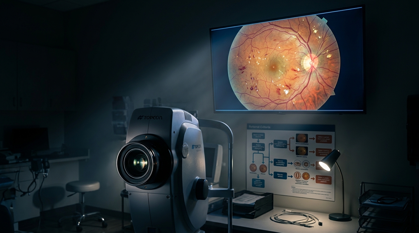
The IDx-DR (now LumineticsCore) was the first FDA-authorized autonomous AI diagnostic system (2018), designed to detect more-than-mild diabetic retinopathy[1] (mtmDR) and diabetic macular edema (DME) from non-mydriatic fundus photography. In the pivotal trial of 900 patients, it achieved sensitivity of 87.2% and specificity of 90.7% for mtmDR[2]. The EyeArt system demonstrated sensitivity of 95.5% and specificity of 86.0% in a real-world validation study of 893 patients across 15 primary care sites[3]. These systems provide results within 60 seconds, eliminating the need for specialist image interpretation.
Severity Classification and Referral Thresholds
The International Clinical Diabetic Retinopathy Severity Scale classifies disease as: no apparent retinopathy, mild nonproliferative DR (NPDR, microaneurysms only), moderate NPDR (more than microaneurysms but less than severe), severe NPDR (20+ intraretinal hemorrhages in each quadrant, venous beading in 2+ quadrants, or intraretinal microvascular abnormalities in 1+ quadrant), and proliferative DR (neovascularization or vitreous/preretinal hemorrhage). Referral to ophthalmology is indicated for moderate NPDR or worse, any DME, or unexplained visual changes.
Treatment Milestones: Anti-VEGF and Laser
Anti-VEGF therapy has replaced panretinal photocoagulation (PRP) as first-line treatment for center-involving DME. The Protocol T trial compared aflibercept, bevacizumab, and ranibizumab: at 2 years, aflibercept produced the greatest visual acuity gain in eyes with baseline VA of 20/50 or worse (18.1 letters versus 13.3 for bevacizumab, p<0.001)[4]. The PANORAMA and RIDE/RISE trials demonstrated anti-VEGF efficacy in preventing progression from severe NPDR to proliferative disease, with a 75% reduction in PDR development at 2 years with aflibercept[5].
Implementing AI Screening in Primary Care
Successful implementation of AI-assisted screening requires a non-mydriatic fundus camera (cost approximately $10,000-$25,000), trained imaging staff (certification typically requires 4-8 hours of training), and a clear referral pathway. Studies from the VA healthcare system and Johns Hopkins demonstrate that point-of-care AI screening increases screening rates from 50% to 80% while reducing time-to-diagnosis by 60%[1]. CMS reimburses autonomous AI screening under CPT 92229 at approximately $40 per eye. The cost-effectiveness ratio is estimated at $8,200 per QALY gained, well below conventional thresholds.
Making Screening Happen: Practical Steps for Your Practice
For the primary care practice or endocrinology clinic considering AI-assisted retinal screening, the implementation path is more straightforward than it might appear. The camera and AI system represent a one-time capital investment that is offset by reimbursement within the first year for most practice volumes. The more significant investment is workflow integration: identifying which staff will operate the camera, building the screening into the diabetes visit workflow so it happens automatically rather than requiring a separate appointment, establishing a direct referral pathway with ophthalmology for positive screens, and tracking completion rates to ensure the program is actually reaching the patients who need it. The practices that achieve the highest screening rates are those that make retinal imaging as routine as checking a hemoglobin A1c — a standard part of every comprehensive diabetes visit rather than an add-on that requires separate scheduling.
Limitations and What AI Screening Does Not Replace
AI screening systems are designed to detect referable diabetic retinopathy — they are not comprehensive eye examinations. They do not assess for glaucoma, cataracts, age-related macular degeneration, or other ocular conditions that a dilated fundoscopic examination would detect. Patients with positive AI screens still require ophthalmologic evaluation for treatment decision-making, and patients with negative screens who report visual symptoms should still be referred. The image quality limitation is real: non-mydriatic photography in patients with small pupils, cataracts, or poor cooperation can produce ungradable images in roughly the same proportion as in-office fundoscopy, necessitating referral for dilated examination. AI screening is a triage tool that dramatically improves access to the first step of care, not a replacement for the specialist relationship that patients with established retinopathy require.
References
- Autonomous AI System for Diabetic Retinopathy in a Primary Care Setting (Abramoff et al., NPJ Digit Med 2019) PubMed 31304320
- IDx-DR Pivotal Trial: AI Autonomous Detection of Diabetic Retinopathy (Abramoff et al., NPJ Digit Med 2018) PubMed 29342388
- Real-World Performance of an Automated AI Screening System for Diabetic Retinopathy (EyeArt) PubMed 34151725
- Aflibercept, Bevacizumab, or Ranibizumab for Diabetic Macular Edema (Protocol T, DRCR.net, NEJM 2015) PubMed 26242766
- PANORAMA: Aflibercept for Nonproliferative Diabetic Retinopathy (Brown et al.) PubMed 34351414
Frequently Asked Questions
What is the sensitivity of IDx-DR for diabetic retinopathy?
When should diabetic retinopathy screening begin?
What is the CPT code for autonomous AI retinal screening?
Which anti-VEGF agent is most effective for diabetic macular edema with poor baseline VA?
What are the referral thresholds for diabetic retinopathy?
Does AI screening improve diabetic retinopathy screening rates?
Explore This Topic in Ailva
Ailva is a free clinical intelligence platform for NPI-verified US physicians. Get evidence-based answers with verified citations from 16M+ indexed papers — plus free CME credits.

Founder of Ailva.ai | Former Director of Research and Author of 200+ Medically Reviewed Articles | Editor-in-Chief of EudaLife Magazine